-
Email info@ijcmcs.org
-
Address 848 N. Rainbow Blvd. #5486 Las Vegas, NV 89107, USA
1Radiology Department, CMH, Peshawar, Pakistan.
2Radiology Department, Agha khan University, Karachi, Pakistan.
*Corresponding author: Nayab Mustansar
Radiology Department, CMH, Peshawar, Pakistan.
Email: drnayabmustansar@gmail.com
Received: Dec 24, 2024
Accepted: Jan 14, 2025
Published Online: Jan 21, 2025
Journal: International Journal of Clinical & Medical Case Studies
Copyright: © Mustansar N (2025). This Article is distributed under the terms of Creative Commons Attribution 4.0 International License
Citation: Mustansar N, Siddiqui CTS, Asghar CA, Shafiq A, Ur Rahman A, et al. Beyond the surface- When anatomy goes awry: A deep dive into gastric anomalies- Insight from a unique case. Int J Clin Med Case Stud. 2025; 2(1): 1005.
We present the case of a 37-year-old female with gastric fullness and early satiety. Imaging revealed a metallic object in the stomach, leading to a Contrast-Enhanced CT scan (CECT) that demonstrated non-rotation of the gut, an annular pancreas, and a duodenal web, resulting in significant duodenal dilatation. The patient also had polysplenia, a left adnexal cyst, and a left renal calculus. Endoscopy retrieved multiple foreign bodies including a plastic button, coin, and thread.
Congenital gastrointestinal anomalies, such as non-rotation of the gut and annular pancreas, can present with a range of symptoms. This case highlights a complex presentation with significant anatomical variations and incidental findings of ingested foreign bodies, emphasizing the need for thorough evaluation and intervention. The interplay between these anomalies can complicate clinical management, as seen in similar reported cases [1].
A 34-year-old female presented to the emergency department of CMH Peshawar with a 4-week history of mild gastric fullness and early satiety. She experienced worsening symptoms but reported no abdominal pain, vomiting, or weight loss. Her medical history was unremarkable with no prior abdominal surgeries.
Physical examination: The patient appeared well-nourished but slightly uncomfortable. Mild epigastric tenderness was noted on examination. Vital signs were stable.
Investigations
CT KUB scan: A non-contrast CT scan revealed a metallic object in the stomach.
Gastroenterology referral: Due to the incidental finding, the patient was referred for further evaluation.
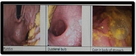
Upper gastrointestinal endoscopy (OGD): OGD revealed several foreign bodies, including a plastic button, coin, and thread. Significant duodenal distension limited visualization as shown in the diagram below:
CECT findings included:
− Non-rotation of the gut.
− Annular pancreas surrounding the duodenum.
− A duodenal web causing a “windsock phenomenon” with second- and third-part duodenal dilatation.
− Polysplenia.
− A left adnexal cyst.
− A left renal calculus.
As shown in figure 2a & b below.
The patient was referred to the surgeon for further work up and her surgery is in plan in the next month (October 2024).

This case presents a rare combination of congenital anomalies and incidental foreign body ingestion, which requires careful analysis to differentiate from other syndromes, such as heterotaxy.
Heterotaxy is characterized by abnormal arrangement of the internal thoracic and abdominal organs and often involves cardiac anomalies. In this patient, polysplenia was noted, which is a specific manifestation of heterotaxy but does not encompass the broader spectrum of conditions that define true heterotaxy. This distinction is crucial as heterotaxy typically presents with more severe structural anomalies, including significant malformations of the heart and lungs, which were absent in this case [2,3].
Complexity of congenital anomalies
Non-rotation occurs during embryonic development, leading to potential complications such as volvulus or obstruction [4,5]. The anatomical mispositioning can complicate surgical approaches and necessitate careful planning.
Annular Pancreas arises when the ventral pancreatic bud encircles the duodenum, which can cause obstructive symptoms [6,7]. In this case, the annular pancreas likely exacerbated the duodenal obstruction from the web.
The presence of a duodenal web leads to proximal obstruction, which can cause significant gastrointestinal distress. The “windsock phenomenon” is a specific radiological finding indicating proximal duodenal dilatation due to this obstruction [8,9]. Recognizing this phenomenon can guide appropriate surgical interventions.
While polysplenia often coexists with other congenital anomalies, its isolated presence in this case highlights the need for comprehensive imaging to assess potential associated conditions [10]. Polysplenia can complicate surgical approaches and increase the risk of splenic complications.
The retrieval of foreign bodies such as a plastic button, coin, and thread suggests accidental ingestion, which can lead to gastrointestinal obstruction or irritation [11]. This case emphasizes the importance of considering foreign body ingestion in patients with unexplained gastrointestinal symptoms, especially in the presence of anatomical anomalies.
Incidental findings
The left adnexal cyst and left renal calculus were incidental findings, yet they warrant monitoring due to their potential clinical significance. Regular follow-up imaging may be necessary to assess for changes in these findings, as they can lead to complications if left untreated.
Management
Surgical intervention is indicated for symptomatic congenital anomalies. Recommended approaches may include:
− Resection of the duodenal web.
− Surgical management of the annular pancreas, potentially involving decompression or resection.
− Evaluation and management of the renal calculus, along with monitoring of the adnexal cyst.
This case underscores the importance of a comprehensive diagnostic approach in patients presenting with nonspecific gastrointestinal symptoms. The complexity of congenital gastrointestinal anomalies, such as non-rotation of the gut, annular pancreas, and duodenal web, requires careful consideration during management. Differentiating between polysplenia and heterotaxy is critical for appropriate surgical planning and patient care. Incidental findings, such as foreign bodies and additional anatomical variations, further complicate the clinical picture, necessitating thorough imaging and endoscopic evaluations. Future studies should focus on the long-term management and outcomes of similar cases to enhance our understanding and treatment strategies for these rare anomalies.