-
Email info@ijcmcs.org
-
Address 848 N. Rainbow Blvd. #5486 Las Vegas, NV 89107, USA
1Department of Neurosurgery, University Medical Center of Limoges, Limoges, France.
2XLIM UMR CNRS 7252 University of Limoges, Limoges, France.
3Department of Neurosurgery, University Medical Center of Henri-Mondor, Créteil, France.
4INSERM U955 Biomedical Research Institute Henri Mondor, Créteil, France.
*Corresponding author: Wassim Khalil
Department of Neurosurgery, University Medical Center of Limoges, Limoges, France.
Email ID: wassim.kh.khalil@gmail.com
Received: Jun 02, 2025
Accepted: Jun 19, 2025
Published Online: Jun 26, 2025
Journal: International Journal of Clinical & Medical Case Studies
Copyright: © Khalil W (2025). This Article is distributed under the terms of Creative Commons Attribution 4.0 International License.
Citation: Khalil W, Apra C, Chirani K, Harimbonona ZM, Ratovoarison FT, et al. Extradural ligamentum flavum cyst: MRI characteristics and surgical management. Int J Clin Med Case Stud. 2025; 2(1): 1023.
Ligamentum Flavum Cysts (LFC) are uncommon and their differentiation from other Juxta-facetal cysts & epidural cystic lesions is difficult based on imaging techniques. We present one such rare case of ligamentum flavum cyst with relevant review of the literature. An eighty-eight years male presented with progressively worsening radicular symptom in the left lower limb. His neurological examination was unremarkable. Magnetic resonance imaging of lumbar spine revealed an epidural cystic lesion narrowing the left lateral recess. Intra-operatively, a mass was found originating from ventral surface of ligamentum flavum. Pathological examination was suggestive of fibrocollagenous tissue without synovial lining. The exact pathogenic mechanism for the formation of LFCs is not well understood. Association with segmental instability and degenerative conditions of spine is postulated. They are commonly seen at the mobile junctional levels of the spine. Persistent micro-traumatic events with abnormal movement maybe contributory to their origin. They present either with radiculopathy or neurogenic claudication symptoms owing to compressive effect on adjacent neural structure. LFC should be considered as a differential in patients with radicular pain or claudication symptoms with epidural cystic lesion seen on MRI. Complete excision of such lesion provides excellent pain relief in symptomatic individuals.
Cysts of the ligamentum flavum of the lumbar spine have seldom been described. They are clearly visible in computed tomography as well as nuclear magnetic resonance, but are frequently wrongly diagnosed as ganglion or synovial cysts. The correct diagnosis is not feasible until after surgery. Such space occupying lesions can most often lead to uniradicular pain due to compression of a root. These cysts should be viewed as part of the degenerative process of the spine but not as tumor lesions. They need to be removed only in case of root entrapment. On the basis of six of our cases treated by surgery we describe the symptoms, imaging findings, operative techniques and pathological investigations.
MRI of a 67-year-old patient presenting with disabling right sided L5 radicular pain without motor deficit.
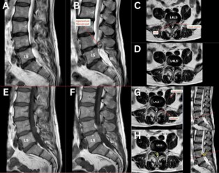
Sagittal (Figure 1A,1B,1E,1F) and axial (Figure 1C,1D,1G,1H) MRI revealed an extradural cystic lesion with a broad base insertion adjacent to the ligamentum flavum at L4/L5, appearing well-demarcated, hyperintense on T2-weighted and hypointense on T1-weighted sequences. The lesion significantly compresses the right L5 root and narrows the spinal canal.
Surgical excision and histology confirmed a fibro-collagenous ligamentum flavum cyst without synovial lining.
Radiological differentiation from ganglion or synovial cysts remains challenging, highlighting the importance of histopathological confirmation [1,2]. Complete cyst excision resulted in resolution of the patient's radicular symptoms.
Disclosure of interest: The authors declare that they have no competing interest.